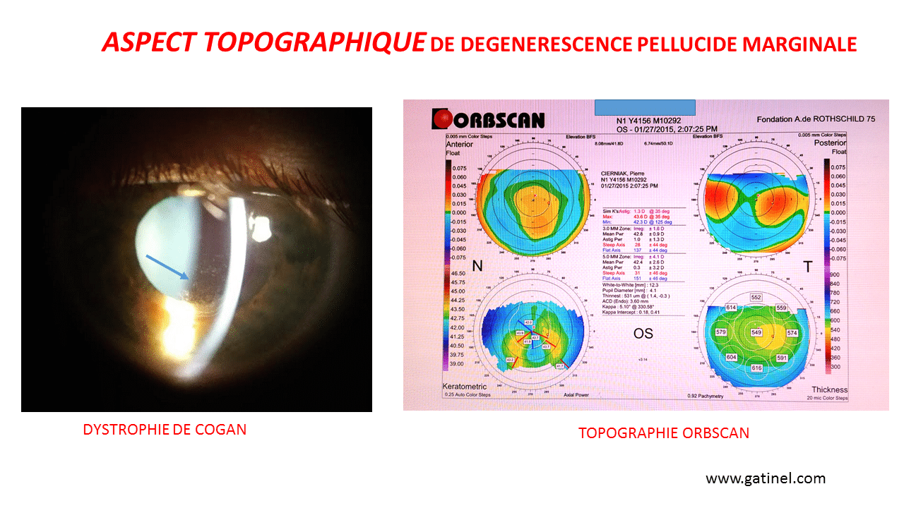


4 Tomography refers to cross-sectional mapping, where the layers of the cornea are seen like layers of a cut cake. It is the ‘gold standard’ in diagnosing and assessing keratoconus. Corneal tomographyĬorneal tomography is a non-invasive way to measure the contours and shape of the cornea. 2 The only way this can be detected reliably is with corneal tomography. According to The Global Consensus on Keratoconus and Ectatic Diseases, for a true diagnosis of keratoconus there must be thinning at the back of the cornea that is not due to another cause, and an irregular corneal profile where some areas are abnormally thin. Your optometrist can detect signs of keratoconus by analysing structural changes in the cornea. Typically, night vision is affected first (haloes and streaks around headlights), which then develops into blurry and distorted vision.

Keratoconus is a multifactorial disease, so the onset and progression can vary significantly between different people, and even between different eyes of the same person. 6 Most studies indicate that keratoconus affects men and women equally, and it is common for each eye to have a differing degree of severity. Keratoconus presents during teenage years and progresses through to your 40s, although the age at which it worsens is variable. 2 Often your optometrist will recommend certain anti-allergy eye drops to decrease symptoms of itchiness and reduce the urge to rub the eyes. 2 Environmental factors include eye rubbing, allergies and hayfever, eczema, asthma and floppy eyelid syndrome. Genetic factors include family history of the disease (especially first-degree relatives) 4, Asian and Arabian ethnicity and congenital conditions like Down, Marfan and Ehlers-Danlos syndromes. 3 The reason why keratoconus develops varies from person to person, where genetic and environmental factors contribute to different degrees. Globally, keratoconus affects 1.3 people in 1000, although in Western countries like Australia these rates double.


 0 kommentar(er)
